angemeldet für: Postervorträge
| Topics: |
01.8 Implantierbare Aggregate
01.5 Therapie Vorhofflimmern - interventionell |
|
*A98*
*A98*
09.01.2018 14:28
|
Herzrhythmusmodell zur Simulation elektrischer und thermischer Felder bei kardialer Resynchronisationstherapie und Hochfrequenz Ablation
M. Krämer1, M. Heinke1
1Electrical Engineering and Information Technology / Medical Engineering, University of Applied Sciences Offenburg, Offenburg;
Hintergrund: Richtung und Stärke des elektrischen Feldes (E-Feld) der biventrikulären (BV) Stimulation und elektrische interventrikuläre Desynchronisation sind bei Patienten mit Herzinsuffizienz und verbreitertem QRS Komplex von Bedeutung für den Erfolg der kardialen Resynchronisationstherapie (CRT). Das 3D Herzrhythmusmodell (HRM) ermöglicht die Simulation von CRT und Hochfrequenz (HF) Ablation. Das Ziel der Studie besteht in der Integration von Schrittmacher- und Ablationselektroden in das HRM zur E-Feld Simulation der BV Stimulation und thermischen Feld (T-Feld) Simulation der HF Ablation von Vorhofflimmern (AF).
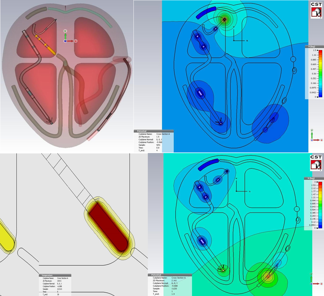
Methoden: Es wurden fünf multipolare linksventrikuläre (LV) Elektroden, eine epikardiale LV Elektrode, vier bipolare rechtsatriale (RA) Elektroden, zwei rechtsventrikuläre (RV) Elektroden und ein HF Ablationskatheter mit CST (Computer Simulation Technology, Darmstadt) modelliert und das HRM (Schalk et al: Clin Res Cardiol 106, Suppl 1, April 2017, P1812) um den Koronarvenensinus (CS) erweitert (HRM-CS). E-Feld Simulationen bei vorhofsynchroner BV Stimulation und bei RA Stimulation mit RV und LV Ableitung erfolgten mit den Elektroden Select Secure 3830, Capsure VDD-2 5038 und Attain OTW 4194 im HRM+CS (Fig.). F-Feld Simulationen der HF Ablation von AF bei CRT wurden mit integriertem Ablationskatheter AlCath G FullCircle (Biotronik) simuliert.
Ergebnisse: HRM-CS ermöglichte 3D E-Feld Simulationen bei vorhofsynchroner bipolarer BV Stimulation und bei bipolarer RA Stimulation mit bipolarer RV und LV Ableitung. RV und LV Stimulation erfolgten zeitgleich bei einer Amplitude von 3 V an der LV Elektrode und 1 V an der RV Elektrode mit einer Impulsbreite von jeweils 0,5 ms. Die von der BV Stimulationen erzeugten Fernpotentiale konnten von der RA Elektrode wahrgenommen werden. Das Fernpotential an der RA Elektrodenspitze betrug 32,86 mV und in 1 mm Abstand von der RA Elektrodenspitze ergab sich ein Fernpotential von 185,97 mV. HRM-CS ermöglichte 3D T-Feld Simulationen der HF Ablation von AF bei CRT. Das T-Feld bei HF Ablation des AV-Knotens wurde mit einer anliegenden Leistung von 5 W bei 420 kHz an der distalen 8 mm Ablationselektrode simuliert. Die Temperatur an der Katheterspitze betrug nach 5 s Ablationsdauer 88,66 °C, in 1 mm Abstand von der Katheterspitze im Myokard 42,17 °C und in 2 mm Abstand 37,49 °C.
Schlussfolgerungen: HRM-CS und Elektrodenmodelle ermöglichen die 3D Simulationen von E-Feldern bei vorhofsynchroner BV Stimulation, RA Stimulation mit RV und LV Wahrnehmung und von T-Feldern bei HF Ablation. E-Feld Simulationen von RA, RV und LV Stimulation und Sensing können möglicherweise zur Vorhersage von CRT Respondern genutzt werden.

Figure 1: Position of the CRT electrode catheters and the ablation catheter in the heart rhythm model (top left), simulation of the potential fields during right atrial pacing with sensing the tips of the right and left ventricular catheter (top right), HF ablation of the AV node after 15 sec (bottom left), simulation of the potential fields during BV pacing with the tips of the right and left ventricular catheter (bottom right).